
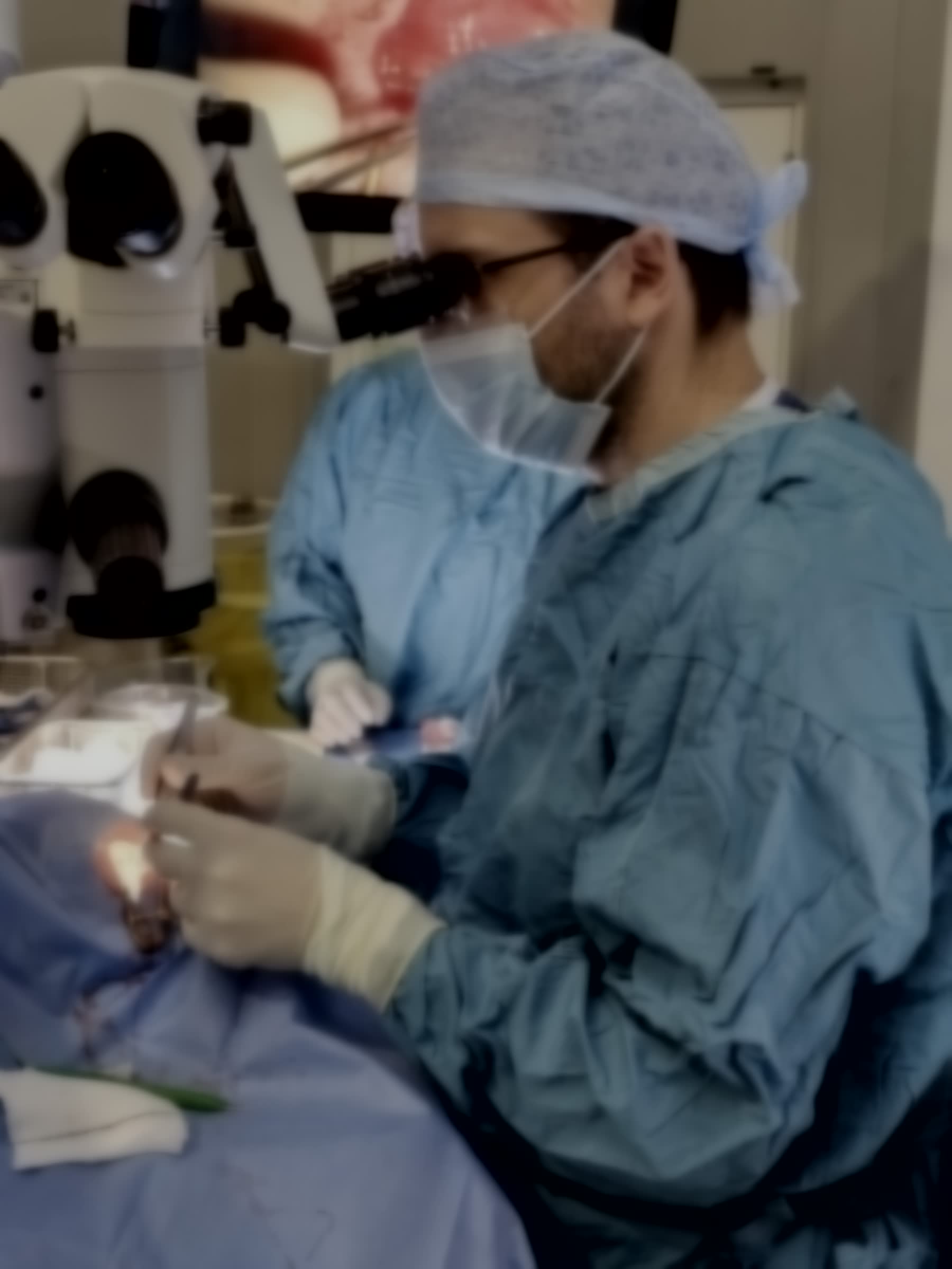
Operating microscope over a sterile surgical field. Fig. 1 · Theatre — emergency repair

RETINA

Procedure · Vitreoretinal surgery

Retinal detachment repair

Surgery to reattach a retina that has peeled away from the back of the eye and to seal the tear that caused it — usually as an emergency, because time is what protects sight.

The aim of the operation is simple to state: close the retinal tear and settle the retina back against the wall of the eye. There are three main techniques, chosen to fit the detachment and often combined.

The first is vitrectomy — keyhole surgery from inside the eye, removing the vitreous and supporting the retina with a gas bubble or, occasionally, silicone oil. The second is a scleral buckle — a soft silicone band stitched onto the outside of the eye to gently indent the wall and support the retina. The third is a pneumatic (gas) technique with laser or freezing (cryotherapy) to seal the tear, used for selected detachments.

Who it is for

Retinal detachment repair is for anyone with a rhegmatogenous (tear-related) retinal detachment. How urgently it is carried out depends on whether the macula — the central reading part of the retina — is still attached: a ‘macula-on’ detachment is more urgent, because operating while the macula is still on is what protects central vision.

A surgeon operating under an ophthalmic microscope in a sterile theatre.
Fig. 1 · Vitreoretinal theatre — sterile field
What to expect

Before, during, and after.

1
Before

Urgent assessment and a plan

Because a detachment is time-critical, assessment and imaging are arranged urgently. The options are explained in plain terms, consent is taken, and a plan is agreed — including any head-posturing you will need afterwards.

2
During

Reattaching the retina

The retina is reattached by the technique chosen for your detachment — vitrectomy, a scleral buckle, gas, or a combination — under local or general anaesthetic. The eye is then filled to hold the retina in place while it heals.

3
After

Healing over weeks

You may need to hold a particular head position for several days, and you must not fly while a gas bubble remains in the eye. Drops guard against infection and inflammation, and vision recovers gradually over weeks. If the macula had detached, central vision may not fully return.

Risks, stated plainly

Reattachment surgery is well established, but no eye operation is without risk. The ones worth understanding are:

  • Re-detachment. The retina can lift away again and need further surgery.
  • Scar tissue. Proliferative vitreoretinopathy — scar tissue on the retina — can form and pull it off again.
  • Cataract. The eye’s natural lens commonly clouds after vitrectomy, and cataract surgery is often needed later.
  • Changes in eye pressure, raised or lowered, after surgery.
  • Double vision, which is more common when a scleral buckle is used.
  • Infection or bleeding inside the eye — uncommon, and treated promptly if it occurs.
  • Limited central vision. Even after successful reattachment, central vision may be limited if the macula was involved.

Your individual risks depend on your eye and the type of detachment, and are discussed honestly before you decide.

Consultation

Talk it through with Dr. Diafas.

Whether retinal detachment repair is right for you is a decision made together, after your scans and symptoms are reviewed.

This page provides general information only and is not a substitute for an individual consultation. If you are worried about your vision, seek medical advice.