
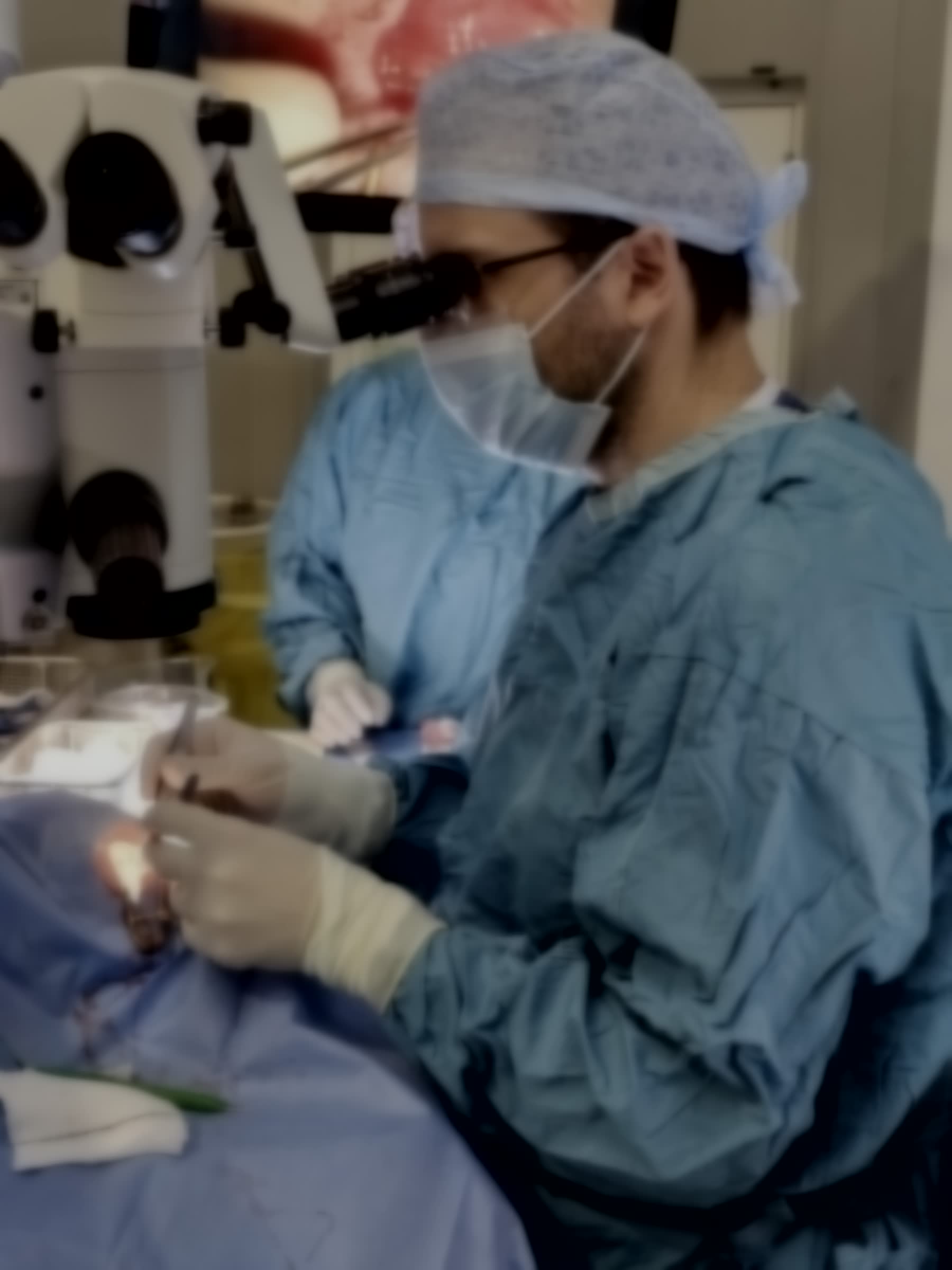
The centre of the eye is filled with a clear jelly called the vitreous. In a vitrectomy, the surgeon makes three tiny openings in the white of the eye — each less than a millimetre — and, working under a microscope, removes that jelly with a fine cutting instrument. With the vitreous out of the way, the retina at the back of the eye can be reached and repaired directly.
It is not one operation but a platform for many: the same access is used to reattach a detached retina, to peel a membrane off the macula, to close a macular hole, to clear blood or scar tissue in diabetic eye disease, and to remove a dislocated lens. At the end, the eye is usually filled with a gas bubble, air, or occasionally silicone oil to support the retina while it heals.
Who it is for
Vitrectomy is considered when a problem sits at the back of the eye and cannot be treated with drops, injections or laser alone. Common reasons include retinal detachment, macular hole, epiretinal membrane, advanced diabetic retinopathy, vitreous haemorrhage, and floaters severe enough to disable vision. Whether it is the right step for you is decided at consultation, on the basis of your scans and your symptoms.